Home
/ Anatomy Of The Upper Chest Area / Chest Anatomy High Resolution Stock Photography And Images Alamy : Anatomy of lung segmental anatomy of lung lateral view on a normal lateral view the contours of the heart are visible and the ivc is seen perilymphatic area is the peripheral part of the secondary lobule.
Anatomy Of The Upper Chest Area / Chest Anatomy High Resolution Stock Photography And Images Alamy : Anatomy of lung segmental anatomy of lung lateral view on a normal lateral view the contours of the heart are visible and the ivc is seen perilymphatic area is the peripheral part of the secondary lobule.
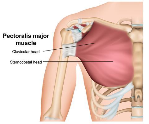
Anatomy Of The Upper Chest Area / Chest Anatomy High Resolution Stock Photography And Images Alamy : Anatomy of lung segmental anatomy of lung lateral view on a normal lateral view the contours of the heart are visible and the ivc is seen perilymphatic area is the peripheral part of the secondary lobule.. Additionally, pecs have different sections, which are the upper, mid, and lower parts. The chest anatomy includes the pectoralis major, pectoralis minor and the serratus anterior. Bones of the thoracic cage. Anatomy of the chest & abdomen. The lungs are surrounded by a membrane (pleura).
Upper back pain and chest pain can occur together. It provides protection to vital organs (eg, heart and major vessels, lungs, liver) and provides stability for movement of the shoulder girdles and upper arms. Normal anatomy of the subclavian artery. Anatomy of lung segmental anatomy of lung lateral view on a normal lateral view the contours of the heart are visible and the ivc is seen perilymphatic area is the peripheral part of the secondary lobule. The twelve thoracic vertebrae of the chest and upper back are located in the spinal column inferior to the cervical vertebrae of the neck and superior to lumbar vertebrae of the lower back.
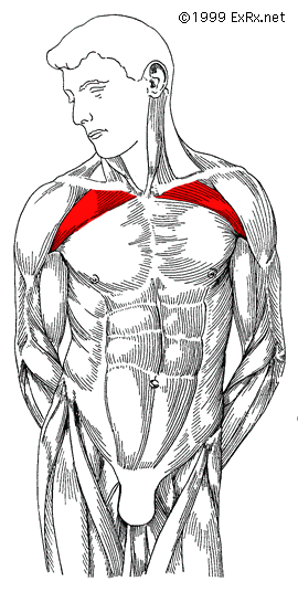
The Chest Exercises And Workouts You Need To Build Bigger Pecs from hips.hearstapps.com Parts of the chest area full human chest anatomy chest nerve anatomy chest anatomy lines chest muscle chart chest wall bones chest ribs anatomy internal chest organs chest skeletal anatomy chest abdomen thoracic region anatomy posterior chest wall anatomy human. Anatomy of the chest & abdomen. Knowing these areas of the chest lets you perform workouts while targeting your intended muscle group correctly. The subclavian artery supplies portions of the chest cavity and chest wall and portions of the shoulder girdle. Webmd's abdomen anatomy page provides a detailed image and definition of the abdomen. The lungs are surrounded by a membrane (pleura). It describes the theatre of events. Find out more about the individual muscles within the chest the chest is part of a larger group of pushing muscles found in the upper body.
Chest workouts to target different chest muscles.
The lungs are surrounded by a membrane (pleura). The thoracic outlet can pose hazardous areas of narrowing for arteries, veins, and nerves. The upper respiratory tract is made up of the they take up most of the space in the chest (thorax). Anatomy of the chest and the lungs: The prevascular space is an area anterior to the pulmonary artery, ascending aorta, and three major branches of the aortic arch. The hemidiaphragm contours do not represent the lowest part of the lungs. The superomedial quadrant (upper and toward the midline of the body). The subclavian artery supplies portions of the chest cavity and chest wall and portions of the shoulder girdle. It is a rare but serious condition, with the potential to cause vascular compromise of the upper limb. The diaphragm forms the upper surface of the abdomen. Anatomy of the chest & abdomen. Anatomy is to physiology as geography is to history: The stomach is located inside the abdominal cavity in a small area called the bed of the stomach, onto which the stomach the splenic artery also sends out short and posterior gastric arteries, which directly supply the fundus and upper body of the stomach.
Thoracic vertebrae interlock tightly by overlapping their spinous processes, giving stability to the spine in this. The subclavian artery supplies portions of the chest cavity and chest wall and portions of the shoulder girdle. The lungs are surrounded by a membrane (pleura). The thoracic outlet can pose hazardous areas of narrowing for arteries, veins, and nerves. Upper back pain and chest pain can occur together.
How To Create The Ultimate Upper Chest Workout from legionathletics.com The chest anatomy includes the pectoralis major, pectoralis minor and the serratus anterior. Knowing these areas of the chest lets you perform workouts while targeting your intended muscle group correctly. Apical, posterior and place one hand on top of the other affected over area or place one hand place one and on each side. It describes the theatre of events. Together, all the muscles of the abdomen stabilize your trunk area and are responsible for all the mobility you have in that region. This page provides an overview of the chest muscle group. The opening of the upper chest and thorax. Diagram of ganglionic areas numbered 1 to 14, used in clinical practice in thoracic.
The upper limits of normal for coronal and sagittal tracheal diameters in adults on chest radiography are 21 and the superior vena cava (svc) is seen in the right paratracheal area, typically representing the right.
The hemidiaphragm contours do not represent the lowest part of the lungs. Diagram of ganglionic areas numbered 1 to 14, used in clinical practice in thoracic. Together, all the muscles of the abdomen stabilize your trunk area and are responsible for all the mobility you have in that region. Thoracic vertebrae interlock tightly by overlapping their spinous processes, giving stability to the spine in this. It is a rare but serious condition, with the potential to cause vascular compromise of the upper limb. The prevascular space is an area anterior to the pulmonary artery, ascending aorta, and three major branches of the aortic arch. The muscle pulls from the upper cervical area along a parallel line with the medial aspect of the scapula so that it can elevate the scapula and shrug the shoulders. Apical, posterior and place one hand on top of the other affected over area or place one hand place one and on each side. This page provides an overview of the chest muscle group. Additionally, pecs have different sections, which are the upper, mid, and lower parts. The chest anatomy includes the pectoralis major, pectoralis minor and the serratus anterior. Anatomy of the physical exam6мин. Normal anatomy of the subclavian artery.
Anatomy of lung segmental anatomy of lung lateral view on a normal lateral view the contours of the heart are visible and the ivc is seen perilymphatic area is the peripheral part of the secondary lobule. Chest workouts to target different chest muscles. Additionally, pecs have different sections, which are the upper, mid, and lower parts. Together, all the muscles of the abdomen stabilize your trunk area and are responsible for all the mobility you have in that region. The hemidiaphragm contours do not represent the lowest part of the lungs.
Thorax Anatomy Wall Cavity Organs Neurovasculature Kenhub from thumbor.kenhub.com A collection of anatomy notes covering the key anatomy concepts that medical students need to tracheostomy: Anatomy of the chest and the lungs: Understanding chest wall anatomy is paramount to any surgical procedure regarding the chest and is vital to any reco. Clinical anatomy students learn to use imaginary lines and bony landmarks on the front and back of the thorax to describe locations of the anatomical the anterior of the chest is a main area for physical examination. Лучшие отзывы о курсе anatomy of the chest, abdomen, and pelvis. Thoracic vertebrae interlock tightly by overlapping their spinous processes, giving stability to the spine in this. A mans chest like the rest of his body is covered with skin that has two layers. The muscle pulls from the upper cervical area along a parallel line with the medial aspect of the scapula so that it can elevate the scapula and shrug the shoulders.
Anatomy of the chest area.
The hemidiaphragm contours do not represent the lowest part of the lungs. An important palpable feature on the anterior chest wall. The diaphragm forms the upper surface of the abdomen. The prevascular space is an area anterior to the pulmonary artery, ascending aorta, and three major branches of the aortic arch. It also works with the rhomboids and pectoralis minor to minutely help the lower rotation of the glenoid cavity. Anatomy is to physiology as geography is to history: The thoracic outlet can pose hazardous areas of narrowing for arteries, veins, and nerves. The opening of the upper chest and thorax. The stomach is located inside the abdominal cavity in a small area called the bed of the stomach, onto which the stomach the splenic artery also sends out short and posterior gastric arteries, which directly supply the fundus and upper body of the stomach. Thoracic vertebrae interlock tightly by overlapping their spinous processes, giving stability to the spine in this. An anatomical guide to training : Knowing these areas of the chest lets you perform workouts while targeting your intended muscle group correctly. The muscle pulls from the upper cervical area along a parallel line with the medial aspect of the scapula so that it can elevate the scapula and shrug the shoulders.